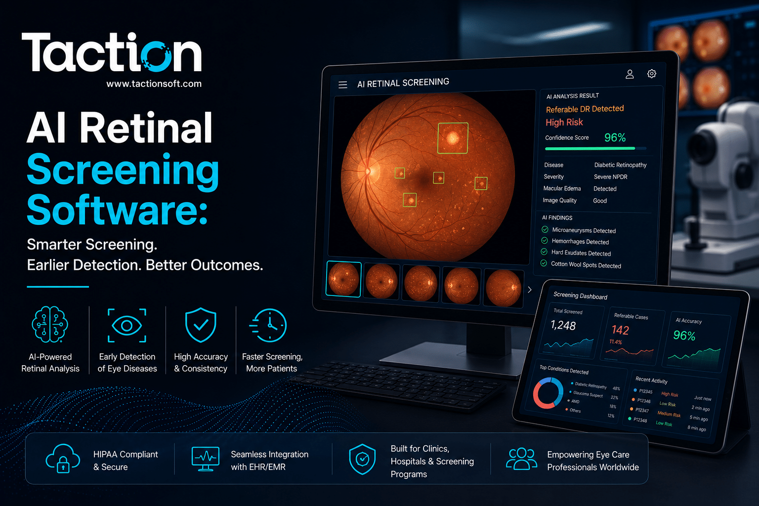
AI retinal screening software analyzes fundus images to detect diabetic retinopathy, macular degeneration, and other retinal disease indicators automatically, without requiring an ophthalmologist to review every image in real time. Diabetic retinopathy screening rates remain persistently low in many primary care and endocrinology settings simply because getting a patient in front of an ophthalmologist for a dilated exam is logistically hard, and AI screening exists specifically to close that access gap by letting a retinal image be captured and read at the point of care. This page explains how automated retinal screening works, where it is deployed clinically today, what the evidence shows on diagnostic accuracy, and what to evaluate before adopting a system.
How Automated Retinal Screening Works
AI retinal screening systems are built around a specific, well-defined image analysis task, which is part of why this category has one of the more mature regulatory track records in medical imaging AI.
Fundus Image Capture and Analysis
A non-mydriatic or mydriatic fundus camera captures a digital image of the retina, which the AI system then analyzes for hemorrhages, microaneurysms, exudates, and other markers of diabetic retinopathy severity, returning a screening result, typically a referral or no-referral recommendation, within minutes.
Autonomous Versus Assistive Screening Models
Some systems are cleared to operate autonomously, providing a screening decision without requiring a specialist to review the image before the result is given to the patient. Others function in an assistive mode, flagging concerning images for a remote ophthalmologist or optometrist to review before finalizing the result. The distinction matters significantly for staffing models and workflow design.
Clinical Evidence and Diagnostic Accuracy
Autonomous AI retinal screening has been studied extensively, partly because diabetic retinopathy screening is a well-bounded clinical task with clear grading standards to validate against.
Sensitivity and Specificity for Diabetic Retinopathy
Multiple prospective studies have demonstrated that autonomous AI systems can achieve sensitivity and specificity for detecting referable diabetic retinopathy comparable to human graders, which is the primary reason regulators have been willing to clear some systems for fully autonomous use without a specialist in the loop.
Performance on Other Retinal Conditions
Detection of diabetic retinopathy is the most mature use case, but many platforms are expanding into age-related macular degeneration and other retinal findings. Evidence for these secondary conditions is generally less extensive than for diabetic retinopathy, so buyers should treat additional disease detection claims with more scrutiny than the core diabetic retinopathy use case.
Where AI Retinal Screening Is Deployed Today
The practical value of this technology shows up most clearly in settings where a dedicated ophthalmology visit is hard to arrange.
Primary Care and Endocrinology Clinics
Placing a retinal camera and screening software directly in a primary care or endocrinology clinic lets patients get their annual diabetic eye exam during a visit they were already attending, rather than being referred out to a separate ophthalmology appointment that many patients never schedule or attend.
Rural and Underserved Care Settings
In areas with limited access to ophthalmology specialists, AI retinal screening combined with a telehealth referral pathway allows a technician to capture the image locally while the interpretation and any necessary specialist follow-up happens remotely, extending screening access without requiring a permanent local specialist presence. This mirrors the broader access model behind our work on remote patient monitoring platform development, where the goal is consistently closing care gaps created by distance and specialist scarcity.
Integration With Diabetes Management Programs
Health systems running structured diabetes management programs increasingly build retinal screening into the annual care pathway alongside other required screenings, since a missed diabetic retinopathy screening is one of the more common gaps in quality measure performance for diabetes care programs.
Evaluating a Retinal Screening System
Before adopting a system, health systems should look closely at both the regulatory clearance and the operational fit with existing clinical staff.
Confirming FDA Clearance for Autonomous Use
If the intended workflow relies on an autonomous result without specialist review, confirm the specific FDA clearance covers autonomous operation, since not every system with strong published accuracy data actually holds clearance for that use case, some are cleared only for assistive use requiring specialist confirmation.
Image Quality and Camera Compatibility
Screening accuracy depends heavily on image quality, and systems validated on one camera manufacturer’s output do not always perform identically with a different camera. Confirm the software is validated against the specific camera hardware your clinic plans to use before purchasing either component separately.
Key Takeaways
AI retinal screening software brings diabetic eye exam access into primary care and endocrinology settings where a dedicated ophthalmology referral often falls through, with the strongest clinical evidence supporting autonomous diabetic retinopathy detection specifically. Before adopting a system, confirm whether the FDA clearance covers autonomous or assistive use, and verify the software’s validation against your specific camera hardware. If your organization is exploring retinal screening as part of a broader diabetes care or population health strategy, talk to our team about your current screening workflow.
Frequently Asked Questions
Can AI retinal screening software operate without an ophthalmologist reviewing the result?
Some systems are FDA cleared for fully autonomous operation for diabetic retinopathy screening specifically, providing a referral or no-referral result without requiring specialist review of every image. Others operate in an assistive mode requiring specialist confirmation.
How accurate is AI retinal screening compared to a human specialist?
Studies on autonomous diabetic retinopathy screening have shown sensitivity and specificity comparable to human graders for referable disease detection, though accuracy for other retinal conditions is generally less extensively validated.
What camera equipment is needed for AI retinal screening?
A non-mydriatic or mydriatic fundus camera is required to capture the retinal image. Screening accuracy is validated against specific camera hardware, so confirm compatibility with the vendor before purchasing.
Can retinal screening software detect conditions other than diabetic retinopathy?
Many platforms are expanding into detecting age-related macular degeneration and other retinal findings, though the clinical evidence base for these additional conditions is generally less mature than for diabetic retinopathy.
Where does AI retinal screening typically get deployed in a health system?
Primary care clinics, endocrinology practices, and rural or underserved care settings are the most common deployment points, since these are the settings where routine ophthalmology referral is hardest to complete consistently.