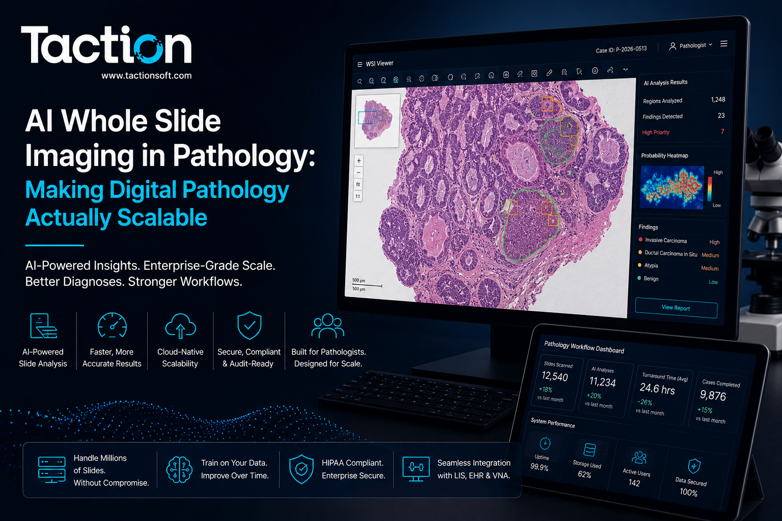
AI whole slide imaging in pathology applies computer vision models directly to the gigapixel-scale digital slides produced by slide scanners, helping pathologists quantify features like mitotic count, tumor cellularity, and biomarker expression across an entire slide rather than relying on manual field-by-field estimation. Whole slide imaging itself solved the problem of digitizing glass slides, but a single scanned slide can be tens of thousands of pixels wide, far more information than a pathologist can exhaustively review manually within a reasonable turnaround time, which is exactly the gap AI analysis is built to close. This page covers how AI models process whole slide images technically, where the clinical value is strongest today, integration into pathology lab workflows, and what to check before adopting a system.
How AI Processes Whole Slide Images
Whole slide images are enormous compared to typical medical images, and the technical approach to analyzing them reflects that scale difference directly.
Tiling and Patch-Based Analysis
Because a whole slide image is far too large to feed into a model in a single pass, most systems break the slide into thousands of smaller image tiles, analyze each tile individually, and then aggregate the tile-level results into a slide-level output, a quantified score, a heatmap, or a region-of-interest annotation the pathologist can review directly on the digital slide.
Weakly Supervised and Multiple Instance Learning
Because pathologists typically label a slide as a whole rather than annotating every individual cell, many whole slide imaging models are trained using multiple instance learning approaches, where the model learns to identify which tiles within a slide are driving an overall diagnosis without requiring exhaustive tile-level labels during training. This training approach is one of the more technically distinctive aspects of pathology AI compared to other imaging AI categories.
Where the Clinical Value Is Strongest
Pathology AI has matured furthest in specific quantitative tasks rather than broad diagnostic replacement.
Quantitative Biomarker Scoring
Biomarkers like Ki-67 proliferation index or HER2 expression require counting stained cells across a slide, a task that is time-consuming and subject to interobserver variability when done manually. AI quantification provides a consistent, reproducible score that reduces variability between pathologists reading the same slide.
Mitotic Figure Counting
Grading certain tumors requires counting mitotic figures across a defined area of the slide, a tedious manual task where AI-assisted counting has shown strong agreement with expert pathologist consensus, freeing up review time for the diagnostic judgment calls that actually require a trained eye.
Prioritization and Triage in High-Volume Labs
In high-volume pathology labs, AI can prescreen slides and flag those with a higher likelihood of malignancy for prioritized review, similar in concept to triage approaches used elsewhere in computer vision for medical imaging, helping labs manage turnaround time on a growing caseload without expanding pathologist headcount at the same rate.
Integrating AI Into the Digital Pathology Workflow
Most pathology labs still operate a hybrid model where glass slides are scanned for specific cases rather than every slide being digitized by default, which shapes how AI tools actually get used day to day.
Compatibility With Slide Scanners and Image Management Systems
AI analysis tools need to ingest whole slide image formats produced by the lab’s specific scanner vendor and integrate with the pathology image management system used to store and retrieve digital slides, since format inconsistencies between scanner vendors remain a real practical barrier to smooth deployment.
Fitting Into the Sign-Out Workflow
The most successful deployments surface AI-generated annotations and scores directly within the pathologist’s existing digital slide viewer at the point of sign-out, rather than requiring a separate application or a disconnected report the pathologist has to cross-reference manually against the slide.
Regulatory Status and Validation Requirements
Pathology AI sits in a regulatory environment that is still maturing relative to radiology AI, and buyers should understand where the clearance landscape currently stands.
FDA Clearance Landscape for Digital Pathology
A growing but still limited number of AI pathology tools hold FDA clearance for specific quantitative tasks, and labs should confirm exactly what a given system is cleared to do, quantification support versus diagnostic assistance carry very different validation standards and liability implications.
Validating Against Your Lab’s Staining and Scanning Protocols
Stain intensity and scanner calibration vary meaningfully between labs, and a model validated on one lab’s staining protocol does not automatically generalize to another lab’s process without local validation. Any pathology AI deployment should include a validation phase against your own historical slides before relying on the tool clinically.
Key Takeaways
AI whole slide imaging analysis has its clearest, best-validated value in quantitative tasks like biomarker scoring and mitotic counting, reducing interobserver variability and freeing pathologist time for diagnostic judgment rather than repetitive counting. Successful deployment depends on scanner and image management system compatibility, integration directly into the sign-out workflow, and local validation against your lab’s specific staining and scanning protocols before clinical use. If your pathology lab is evaluating this technology, get in touch with our team to discuss your current digital pathology infrastructure.
Frequently Asked Questions
Does AI whole slide imaging replace the pathologist’s diagnosis?
No. Current clinically deployed tools are designed to support quantification tasks and prioritization, with the pathologist making the final diagnostic determination based on the full slide review.
What image formats do AI pathology tools support?
Support depends on the specific vendor and typically covers whole slide image formats from major scanner manufacturers, though format compatibility should be confirmed against your lab’s specific scanner before purchasing.
Is AI whole slide imaging analysis FDA cleared?
Some tools have received FDA clearance for specific quantitative tasks such as biomarker scoring. Clearance scope varies significantly by vendor and use case, so confirm exactly what a given system is cleared to perform.
How does AI analysis handle differences in staining between labs?
Performance can vary based on staining protocol and scanner calibration differences between labs. Local validation against your own historical slides before clinical use is strongly recommended regardless of vendor-reported external validation results.
What is the most established clinical use case for pathology AI today?
Quantitative biomarker scoring, such as Ki-67 proliferation index and HER2 expression assessment, along with mitotic figure counting, currently have the strongest published clinical evidence and regulatory track record among pathology AI applications.