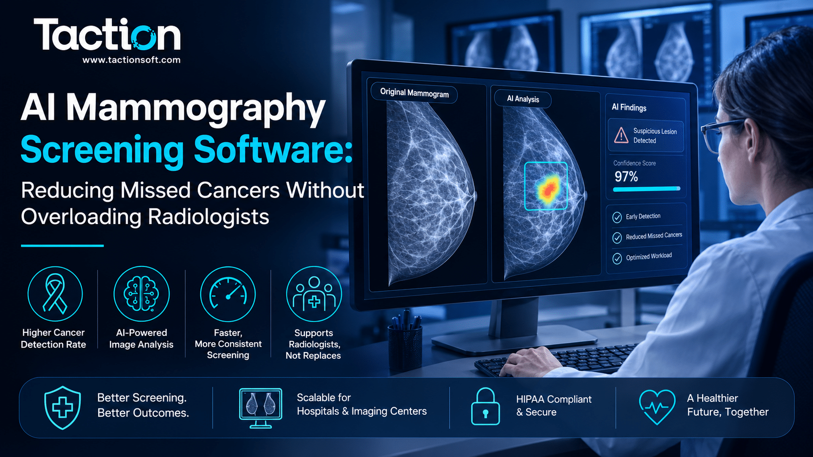
AI mammography screening software analyzes digital breast images to flag regions suspicious for malignancy, acting as a second reader alongside the radiologist to catch findings that are easy to overlook, particularly in dense breast tissue where masking effects reduce visual sensitivity. Breast screening programs read enormous volumes of largely normal studies, and the challenge has never been a shortage of imaging capacity, it has been sustaining consistent attention across thousands of negative reads to catch the rare abnormal one. This page explains how AI screening tools function within existing mammography workflows, what the evidence shows on detection and workload, key considerations around dense breast tissue, and how to evaluate a system before deployment.
How AI Screening Software Fits Into the Mammography Workflow
AI mammography tools generally operate in one of two configurations, both designed to work alongside, not instead of, the reading radiologist.
Concurrent Reader Support
In concurrent mode, the AI system provides its assessment at the same time the radiologist reviews the study, often as a risk score or heatmap overlay indicating regions of concern. The radiologist sees both the raw images and the AI output together before finalizing the report.
Triage and Prioritization Mode
Some deployments use AI purely for worklist triage, scoring studies by likelihood of abnormality so that higher-risk exams are read earlier in the day when radiologist attention is freshest, while lower-risk studies are read later or, in some regulatory frameworks, with reduced double-reading requirements.
What the Evidence Shows on Detection and Workload
Mammography AI has one of the larger evidence bases in medical imaging AI, partly because national screening programs in several countries have run large prospective studies.
Cancer Detection Rate Impact
Large-scale studies, including national screening trials, have shown AI-assisted reading can match or modestly improve cancer detection rates compared to standard double reading, while in some configurations reducing the number of radiologist reads required per study. The specific gain depends heavily on the population screened and the baseline recall rate of the reading radiologists involved.
False Positive and Recall Rate Considerations
A poorly calibrated system increases recall rates and biopsy referrals without a proportional gain in cancer detection, which drives unnecessary patient anxiety and cost. Any deployment decision should weigh the vendor’s reported specificity data as carefully as its sensitivity claims, not just the headline detection number.
Dense Breast Tissue and Masking Effects
Dense fibroglandular tissue appears white on a mammogram, the same as many cancers, which visually masks tumors and is one of the primary reasons screening mammography has known sensitivity limitations in a meaningful share of the screened population.
Where AI Adds the Most Value
AI models trained specifically on dense tissue cases can identify subtle density irregularities and architectural distortion that are statistically associated with malignancy but difficult for the human eye to consistently flag across a busy reading day. This is one of the clearest use cases where the assistive value of the technology is well supported by published research, rather than a marginal efficiency gain.
Combining AI With Supplemental Screening
For patients with dense breasts already receiving supplemental ultrasound or MRI screening, AI mammography output should be treated as one input alongside those modalities, not a substitute for the supplemental imaging protocol already recommended by current screening guidelines.
Integration and Regulatory Considerations
Deploying mammography AI requires connecting it into the existing PACS and reporting pipeline used by the breast imaging center, and confirming its regulatory clearance matches the intended use.
PACS and Reporting Integration
The system needs to ingest full-field digital mammography or tomosynthesis studies directly from PACS and return its output in a format the radiologist’s reporting software can display alongside the images, without requiring a separate login or a disconnected review step that adds friction to an already high-volume workflow. This mirrors the broader integration challenges covered in our page on computer vision for medical imaging, where workflow fit determines whether a technically strong model actually gets used.
FDA Clearance and Intended Use Statement
Confirm the FDA clearance explicitly covers the imaging modality in use, full-field digital mammography versus tomosynthesis are cleared separately in many cases, and that the intended use statement matches how you plan to deploy it, concurrent reading, triage, or standalone reading in jurisdictions where that is permitted.
Planning a Pilot Deployment
Breast imaging centers considering AI screening software should start with a retrospective validation run against a sample of their own historical studies before going live, since vendor-reported performance on external datasets does not always transfer cleanly to a different patient population, equipment vendor, or reading pattern. Centers scoping the investment alongside other imaging AI initiatives can review our detailed breakdown of medical imaging AI development costs to understand how mammography-specific projects compare in scope and cost.
Key Takeaways
AI mammography screening software works best as a concurrent second reader or a triage tool layered into existing PACS workflows, with the strongest evidence supporting its value in dense breast tissue cases where masking effects reduce standard visual sensitivity. Before deploying, confirm FDA clearance matches your specific modality and use case, and run a retrospective validation on your own study population rather than relying solely on vendor benchmarks. If you are evaluating this technology for your breast imaging program, connect with our team to discuss your current PACS and reading volume.
Frequently Asked Questions
Does AI mammography software replace the second radiologist reader?
In most current regulatory frameworks, no. AI is used alongside human readers as a concurrent aid or triage tool. Some jurisdictions have piloted AI as a standalone first reader, but this is not yet standard practice.
How does AI mammography software handle dense breast tissue?
Models trained specifically on dense tissue cases can flag subtle density irregularities that are statistically linked to malignancy but harder to visually distinguish from normal dense tissue, which is one of the clearest documented benefits of the technology.
Will AI mammography software increase our recall rate?
It depends on how the system is calibrated. A well-validated system should not meaningfully increase false positive recalls, but poorly calibrated systems can drive unnecessary callbacks, which is why reviewing specificity data, not just sensitivity, matters before adoption.
What imaging modalities does AI mammography software support?
Support varies by vendor and FDA clearance status. Full-field digital mammography and digital breast tomosynthesis are often cleared separately, so confirm the specific modality coverage before purchasing.
How long does it take to validate an AI mammography system before full deployment?
A retrospective validation run against your own historical study population typically takes four to eight weeks, depending on data access and the size of the validation sample used.